
A milestone for neuroscience: researchers from Oldenburg and London have created the first-ever 3D atlas of a migratory bird’s brain.
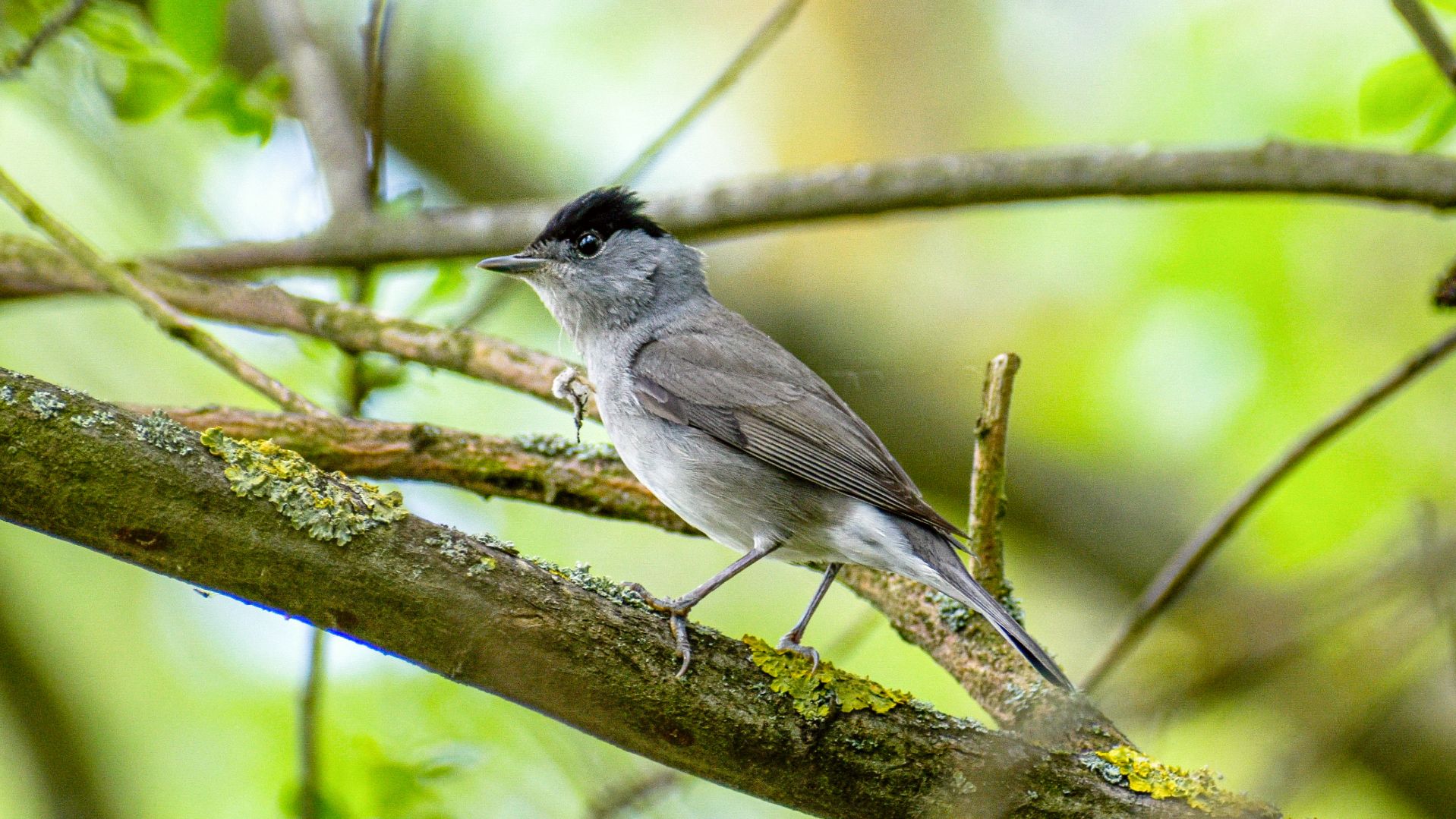
Migrating birds display remarkable navigational capabilities, often reaching centimetre precision on their biannual journeys between wintering and breeding grounds. While neuroscience has only started to unveil the multimodal sensory foundations underlying navigation, a team of researchers from the new Cluster of Excellence at University Oldenburg together with researchers from the Sainsbury Wellcome Centre at UCL London, UK, has now generated the first digital, open-source 3D brain atlas of a migratory bird, the Eurasian blackcap (Sylvia atricapilla). They used high-resolution light microscopy, developed open-source software tools, and published the detailed processes needed to generate new brain atlases for any species. The new atlas will be a valuable resource for neuroscience worldwide and was published in Current Biology.
“The 3D renderings were mind-blowing, allowing you to take a virtual walk through a bird brain at neuron scale. Being able to see neuronal structures and connectivities from any angle does not only look cool, but will significantly accelerate our understanding of the neuronal mechanisms underlying navigation”, said neuroanatomist and Senior Researcher Dr. Dominik Heyers, who held the scientific lead at University of Oldenburg.
Brain atlases – digital, high-resolution, 3D maps of brain structures – are transforming neuroscience. They improve the ability of researchers to interpret their own data, enable cross-validation between and within experiments, and facilitate consensus on the characterization of specific brain areas to foster collaboration. In this publication, the team has revealed a previously unknown link between magnetosensitive areas in the brain and the decision-making “prefrontal” caudolateral nidopallium (NCL), thereby demonstrating how the atlas assists in characterizing novel brain pathways. The published atlas will be regularly updated to incorporate any new data – driving forward our knowledge on learning, memory and cognition.
Speaking the same language when it comes to the brain
“A digital open-source brain atlas allows researchers to directly align their own experimental multimodal data to the common coordinate space of the atlas. It enables consistency, meaning researchers around the world can speak the same language when it comes to the brain. We are delighted to bring this resource to the community, and even more excited about building many more atlases for other research communities in the future,” said Dr. Simon Weiler, Senior Research Fellow at the Sainsbury Wellcome Centre at UCL, and co-lead author of the study.
To create the atlas, the team at SWC used serial two-photon (STP) tomography to image eight male Eurasian blackcap brains. This advanced imaging technique results in well-aligned 2 x 2 x 5 μm voxel size images of entire brains. The individual 3D images from different brains were then iteratively aligned and averaged to create a representative brain template. Following this, experts at University of Oldenburg manually annotated the template, resulting in 44 segmented brain areas, including principal brain compartments, prominent anatomical subdivisions, the song system, and sensory regions implicated in magnetic field processing. Finally, the atlas was incorporated into the BrainGlobe ecosystem and automatic registration, cell detection and object mapping were demonstrated on experimental data.
“To me, this is a key tool that the migration, navigation, and magnetoreception community has been lacking for decades. It will greatly improve consistency and comparability between studies,” said Prof. Dr. Henrik Mouritsen, University of Oldenburg, a co-author of the study.
Even historic samples can be mapped onto the atlas
The new Eurasian blackcap atlas is freely accessible via BrainGlobe for the neuroscience research community and will advance studies in a wide range of topics. The technology allows both new and any yet existing brains, even historic histological samples that have been stored for years on glass slides, to be mapped onto the atlas.
“The core aim of BrainGlobe is to democratise computational neuroanatomy. Creating novel atlases is a step in achieving this. All parts of the pipeline are open-source, and over the coming months we will be improving it so that we, and anyone else, can rapidly create new atlases,” said Dr Adam Tyson, Head of the Neuroinformatics Unit at the Sainsbury Wellcome Centre at UCL and lead of the BrainGlobe Initiative.
The team is currently working on creating a digital 3D brain atlas of the zebra finch (Taeniopygia guttata), a bird species widely used in vocal learning research.
This research was generously funded by the Deutsche Forschungsgemeinschaft (SFB 1372 and EXC 3051), the European Research Council, the Chan Zuckerberg Initiative DAF, the Gatsby Charitable Foundation, Wellcome, and the Alexander von Humboldt Foundation.
This text is based on a press release by the Sainsbury Wellcome Centre.